
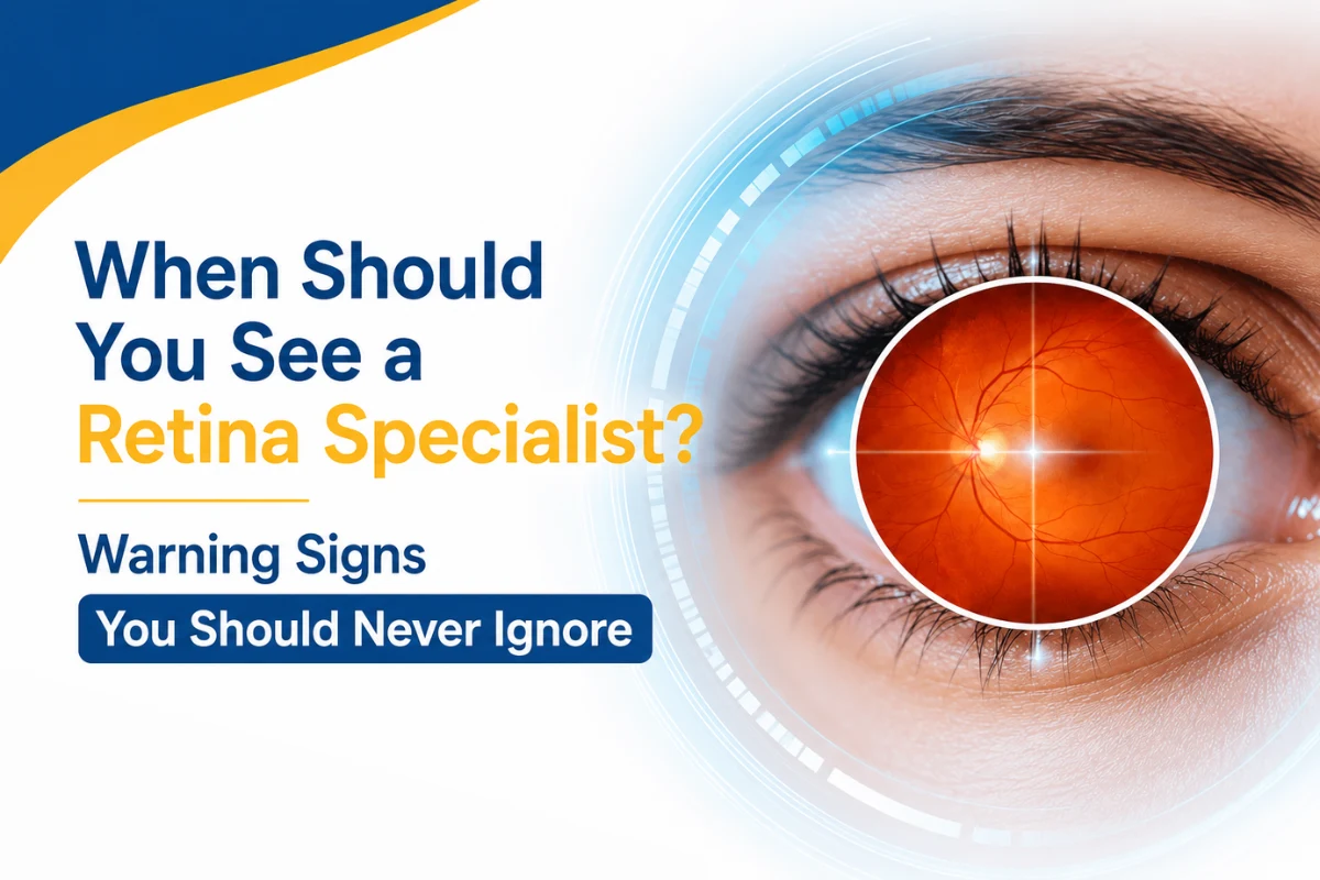
A sudden vision change can be frightening, especially when it appears without pain or redness. You may notice flashing lights, several new floaters, a shadow in one corner of your sight or straight lines beginning to look wavy.
These symptoms should never be dismissed as tiredness or a simple change in spectacle power. Some retinal conditions progress quickly, and delaying an eye examination may increase the risk of permanent vision loss.
The retina is a delicate layer of light-sensitive tissue at the back of the eye. It receives light and converts it into signals that travel through the optic nerve to the brain. When the retina is torn, detached, swollen, bleeding or affected by abnormal blood vessels, vision can change suddenly or gradually.
A retina specialist diagnoses and manages conditions involving the retina and vitreous. These include retinal tears and detachments, diabetic eye disease, age-related macular degeneration, retinal vein occlusion, macular swelling and other conditions affecting the back of the eye. Retina specialists may provide medical treatments such as eye injections and laser procedures, as well as surgery when required.
Patients searching for the best retina specialist in Dubai should first understand which symptoms require immediate assessment and which retinal conditions may remain silent until significant damage has developed.
What Does the Retina Do?
The retina works much like the sensor inside a camera. It captures the light entering the eye and helps create the images that you see.
The central part of the retina contains an area called the macula. The macula is responsible for detailed, straight-ahead vision. You depend on it when you:
- Read
- Recognise faces
- Drive
- Use a mobile phone
- Work on a computer
- See colours and fine details
- Perform close-up tasks
The outer areas of the retina support peripheral or side vision. Problems affecting different parts of the retina can therefore produce different symptoms.
A condition affecting the macula may make words appear blurred or straight lines look bent. A retinal tear or detachment may cause flashing lights, sudden floaters or a curtain-like shadow. Retinal bleeding can produce dark spots or cloudy vision.
What Is a Retina Specialist?
A retina specialist is an ophthalmologist who has additional training in diagnosing and treating diseases of the retina and vitreous.
The vitreous is the clear, gel-like substance that fills the large space between the lens and the retina. Changes in the vitreous can sometimes pull on the retina and create a retinal tear.
A retina specialist may manage conditions such as:
- Retinal tears
- Retinal detachment
- Diabetic retinopathy
- Diabetic macular oedema
- Age-related macular degeneration
- Retinal vein occlusion
- Macular holes
- Epiretinal membranes or macular puckers
- Vitreous haemorrhage
- Retinal complications after eye surgery
- Retinal trauma
- Inherited retinal conditions
Not every visual problem requires retinal treatment. However, when a retinal condition is suspected, a detailed examination of the back of the eye can help identify whether observation, injections, laser treatment or surgery is needed.
When Is a Retinal Symptom an Emergency?
Some retinal symptoms require assessment on the same day. This is particularly important when the change is sudden, affects one eye or is accompanied by a shadow or loss of part of the visual field.
Seek urgent eye care if you notice:
- A sudden increase in floaters
- Repeated flashes of light
- A dark curtain or shadow in your vision
- Sudden loss of part or all of the vision in one eye
- Sudden severe blurring
- Distorted central vision that appears suddenly
- New symptoms after an eye injury
- Sudden vision changes after recent eye surgery
Do not wait to see whether these symptoms improve by themselves. Retinal conditions are not always painful, and the absence of discomfort does not mean that the eye is safe.
Warning Sign 1: A Sudden Increase in Floaters
Floaters are small shapes that appear to move across your field of vision. They may look like:
- Black dots
- Tiny flies
- Threads
- Cobwebs
- Rings
- Squiggly lines
- Clouds or shadows
A few longstanding floaters are common, particularly as people grow older. They often become more noticeable against a bright background, such as a white wall or clear sky.
The concern is not simply whether you have floaters. The concern is whether several new floaters have appeared suddenly.
A sudden shower of dots, dark spots or cobweb-like shapes may indicate that the vitreous gel has pulled away from the retina. This is called a posterior vitreous detachment.
Most vitreous detachments do not require treatment. However, the movement of the vitreous can sometimes pull strongly enough to tear the retina. A retinal tear may then allow fluid to travel underneath the retina and cause a retinal detachment.
Arrange an urgent dilated retinal examination if:
- You suddenly see many new floaters
- The floaters are accompanied by flashes
- Your vision becomes cloudy
- A dark area appears in your peripheral vision
- You have recently had eye surgery or an eye injury
An examination is the only reliable way to determine whether the symptoms are due to a harmless vitreous change or a retinal tear.
Warning Sign 2: Flashes of Light
Retinal flashes are often described as:
- Lightning streaks
- Camera flashes
- Flickering lights
- Sparks
- Bright arcs
- Stars at the edge of vision
They are often more noticeable in the dark or when moving the eyes.
Flashes may occur when the vitreous pulls or rubs against the retina. While this can happen during a routine vitreous detachment, it may also be associated with a retinal tear.
New flashes combined with several new floaters require urgent assessment. The National Eye Institute and the American Academy of Ophthalmology identify sudden flashes and floaters as possible signs of a torn or detached retina.
Not every flash comes from the retina. Migraine-related visual symptoms may create shimmering or zigzag patterns, often involving both eyes. However, patients should not try to diagnose the cause themselves, particularly when the symptom is new, affects one eye or is associated with floaters.
Warning Sign 3: A Dark Curtain, Shadow or Missing Area of Vision
A dark curtain or shadow moving across the vision is one of the most important warning signs of retinal detachment.
The shadow may begin:
- At the side of the vision
- From above or below
- As a dark corner
- As a missing section of the visual field
- As a grey curtain spreading towards the centre
Retinal detachment occurs when the retina pulls away from its normal position at the back of the eye. The detached part of the retina cannot function normally and may lose its blood supply.
The condition is usually painless. A patient may therefore delay seeking help because the eye does not feel uncomfortable.
A retinal detachment is an emergency. A sudden increase in floaters, flashes of light and a curtain-like shadow are its recognised warning symptoms. A dilated eye examination is needed to confirm the diagnosis, and treatment may involve laser or retinal surgery.
Do not drive yourself if a large part of your vision is missing. Arrange safe transportation and seek urgent ophthalmic care.
Warning Sign 4: Sudden, Painless Blurring or Vision Loss in One Eye
Sudden painless vision loss should never be ignored.
A patient may wake up with blurred vision in one eye or notice that part of the image has suddenly become dark, foggy or unclear. This can be caused by several conditions, including:
- Retinal detachment
- Retinal vein occlusion
- Bleeding into the vitreous
- Diabetic retinal bleeding
- Macular disease
- A problem affecting blood flow to the retina or optic nerve
Central retinal vein occlusion occurs when the main vein carrying blood away from the retina becomes blocked. It commonly causes blurred vision or sudden vision loss, usually in one eye. Early diagnosis and treatment can help reduce the risk of further visual damage.
Sudden visual loss can also have neurological or vascular causes. It is not possible to identify the cause safely without medical assessment.
Seek urgent care even if:
- There is no pain
- The eye is not red
- The vision improves slightly
- You have experienced a similar episode before
- You believe your blood sugar or blood pressure caused the problem
Warning Sign 5: Straight Lines Begin to Look Wavy
Distorted vision is known medically as metamorphopsia.
You may notice that:
- Door frames look bent
- Window grills appear curved
- Lines of text look wavy
- Tiles appear uneven
- Faces look distorted
- Objects seem larger or smaller in one eye
- The centre of an image appears pulled or stretched
These changes may indicate a problem affecting the macula.
Possible causes include:
- Wet age-related macular degeneration
- Macular oedema
- Diabetic macular oedema
- Epiretinal membrane
- Macular hole
- Retinal vein occlusion
Age-related macular degeneration affects the macula and can cause blurry or wavy areas in central vision. Early AMD may produce no noticeable symptoms, while later disease can make reading, driving and recognising faces more difficult.
Cover one eye and then the other when checking the symptom. A problem affecting only one eye may be missed when both eyes remain open because the stronger eye can compensate.
New or rapidly worsening distortion should be assessed promptly.
Warning Sign 6: A Blurred, Dark or Blank Spot in Central Vision
A central blind spot can make it difficult to see the object you are directly looking at.
Patients may describe:
- A grey area over words
- Missing letters while reading
- A blurred area in the centre
- Difficulty seeing facial features
- A dark patch over the point of focus
- A blank space in an otherwise clear image
This may be associated with macular degeneration, macular swelling, a macular hole or another condition affecting central retinal vision.
Macular oedema occurs when damaged or abnormal blood vessels leak fluid into the macula, causing it to swell. Symptoms can include blurry or wavy vision and colours appearing dull or washed out.
Do not assume that a central blurred spot is caused by cataract or a change in glasses. Cataract usually affects the clarity of the overall image, while macular disease may create distortion or a more localised central area of missing vision.
Warning Sign 7: Vision Changes When You Have Diabetes
People with diabetes should not wait for visual symptoms before having their retina examined.
Diabetic retinopathy develops when prolonged high blood sugar damages the small blood vessels in the retina. In its early stages, it may cause no symptoms. A patient may see clearly while retinal changes are already present.
As the condition progresses, symptoms may include:
- Blurred vision
- Fluctuating vision
- Dark floating spots
- Difficulty seeing at night
- Faded colours
- Areas of missing vision
- Sudden cloudiness caused by bleeding
Diabetic macular oedema can develop when damaged blood vessels leak fluid into the macula. This can blur central vision and interfere with reading, driving and recognising faces.
The National Eye Institute states that early diabetic retinopathy frequently has no symptoms. Later disease may cause blurred vision, floating spots and serious vision loss. A comprehensive dilated eye examination is therefore essential for early detection.
A retinal assessment is particularly important if:
- You have recently been diagnosed with diabetes
- Your blood sugar has been difficult to control
- You have high blood pressure or kidney disease
- You are pregnant and have pre-existing diabetes
- You have already been diagnosed with diabetic retinopathy
- Your vision has suddenly become blurred
- You see new floaters or dark spots
Good glucose and blood-pressure control remain important, but they do not replace a retinal examination.
Warning Sign 8: A Sudden Cloud, Haze or Red Tint in the Vision
Bleeding into the vitreous cavity is called a vitreous haemorrhage.
A patient may describe:
- A red or brown tint
- Dark clouds
- Cobweb-like shadows
- Hazy vision
- Numerous black spots
- Sudden severe blurring
- A shadow that moves with the eye
Possible causes include diabetic retinopathy, retinal tears, retinal detachment, retinal vein occlusion and trauma.
The bleeding may be mild, or it may make it difficult to see shapes and movement. Even if some of the blood settles and the vision improves, the underlying cause may still require treatment.
Do not assume that the blood will simply clear on its own. A retinal tear or abnormal blood vessel may continue to bleed.
Warning Sign 9: Vision Changes After Eye Surgery
New floaters, flashes, distortion or reduced vision after eye surgery should be reported promptly.
Some temporary blurring may be expected during recovery from certain procedures. However, sudden or worsening symptoms can indicate:
- Retinal tear or detachment
- Retinal swelling
- Infection
- Bleeding
- Changes in eye pressure
- Movement of an implanted lens
- Inflammation
Cataract surgery patients should especially report flashes, a sudden increase in floaters or a curtain-like shadow. Previous eye surgery can be relevant when assessing retinal risk.
Follow the postoperative emergency instructions provided by the surgeon rather than waiting for the next scheduled appointment.
Warning Sign 10: Vision Changes After an Eye Injury
Blunt or penetrating eye injuries can damage the retina even when the front of the eye appears normal.
Seek urgent assessment after eye trauma if you experience:
- Reduced vision
- Flashes or floaters
- A shadow in the visual field
- Double vision
- Blood inside the eye
- Severe pain
- Sensitivity to light
- Distortion
- Difficulty moving the eye
Retinal tears and detachments may develop immediately or sometime after an injury. Always tell the ophthalmologist about previous sports injuries, workplace accidents, falls or road traffic accidents involving the eye or head.
Who Should Have Regular Retinal Examinations?
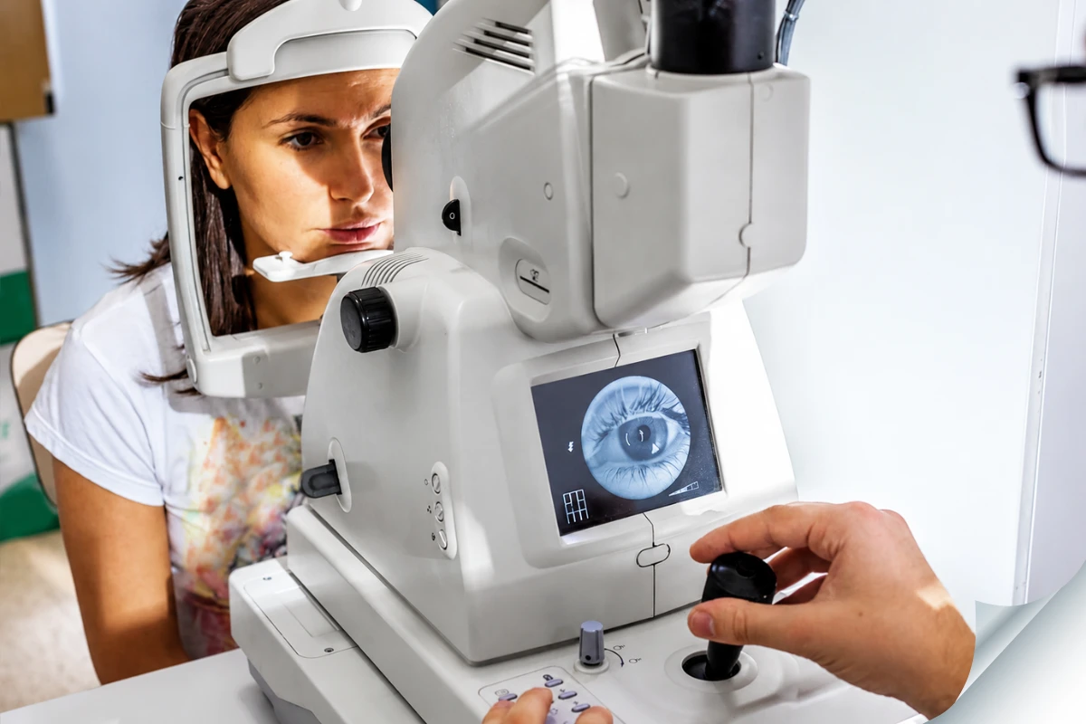
You may need regular retinal monitoring even when you have no symptoms.
Discuss the appropriate examination schedule with your ophthalmologist if you have:
- Diabetes
- Age-related macular degeneration
- High short-sightedness
- Previous retinal tear or detachment
- Retinal disease in the other eye
- A family history of inherited retinal disease
- Previous eye injury
- Previous eye surgery
- Retinal vein occlusion
- High blood pressure with visual symptoms
- Unexplained reduction in vision
- Abnormal findings during a routine eye examination
Patients with diabetes and early macular disease may feel that their vision is normal. Screening allows retinal changes to be detected before they begin significantly affecting daily life.
What Happens During a Retina Examination?
A retinal consultation usually begins with questions about:
- When the symptoms started
- Whether one or both eyes are affected
- Whether the change was sudden or gradual
- Previous eye surgery or injury
- Diabetes and blood pressure
- Current medicines
- Previous retinal conditions
- Family eye history
The specialist may then perform several tests.
Dilated retinal examination
Eye drops are used to widen the pupils. This allows the doctor to examine the retina, macula, blood vessels and optic nerve.
Your near vision may be blurred, and your eyes may be sensitive to light for several hours after dilation. It may be safer to arrange transportation rather than driving yourself.
Optical coherence tomography
Optical coherence tomography, or OCT, creates detailed cross-sectional images of the retina.
It can help detect:
- Macular swelling
- Fluid beneath the retina
- Macular holes
- Epiretinal membranes
- Changes caused by AMD
- Diabetic macular oedema
Retinal photography
Special cameras may be used to document the appearance of the retina. The images can be compared during future appointments.
Fluorescein angiography
A dye is injected into a vein, usually in the arm, and photographs are taken as it passes through the retinal blood vessels.
This can help identify leakage, blocked blood flow and abnormal blood vessels.
Ultrasound scanning
When bleeding, cataract or another obstruction prevents a clear view of the retina, ultrasound may help assess whether a retinal detachment or another structural problem is present.
Visual acuity and eye-pressure testing
The doctor may also check vision, spectacle prescription and eye pressure as part of the complete assessment.
What Treatments Can a Retina Specialist Provide?
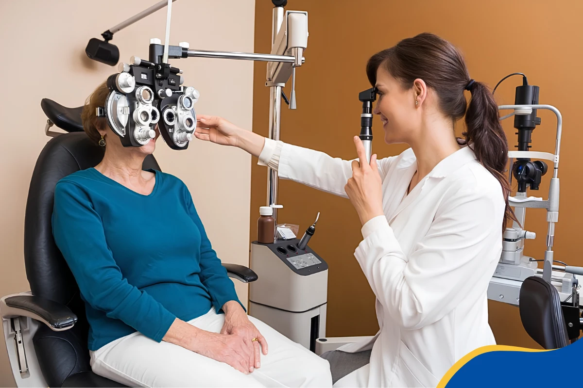
Treatment depends on the diagnosis, severity and part of the retina involved.
Possible treatments include:
Observation and monitoring
Some conditions, such as an uncomplicated vitreous detachment or mild epiretinal membrane, may initially require observation.
Monitoring does not mean that the problem is being ignored. Follow-up allows the doctor to check for progression.
Retinal laser treatment
Laser treatment may be used to seal selected retinal tears, treat abnormal blood vessels or manage certain stages of diabetic retinopathy.
Eye injections
Anti-VEGF or steroid injections may be used for conditions such as:
- Wet age-related macular degeneration
- Diabetic macular oedema
- Retinal vein occlusion
- Other conditions involving retinal swelling or abnormal blood vessels
Anti-VEGF medicines reduce the activity of a protein that promotes blood-vessel leakage and abnormal vessel growth.
Retinal surgery
Surgery may be required for:
- Retinal detachment
- Persistent vitreous haemorrhage
- Macular hole
- Significant epiretinal membrane
- Certain complications of diabetic retinopathy
- Retained lens material or other surgical complications
The specialist should explain the goal of treatment, expected recovery, possible risks and whether additional procedures may be required.
How to Choose the Best Retina Specialist in Dubai
The word “best” is subjective. No doctor can be considered the right specialist for every patient or every retinal condition.
When searching for the best retina specialist in Dubai for your needs, consider:
- The ophthalmologist’s qualifications and current professional licence
- Additional training in medical or surgical retina
- Experience treating your particular condition
- Access to OCT, retinal imaging, angiography and ultrasound
- Availability of laser, injections and surgical care where required
- Clear explanation of the diagnosis
- Honest discussion of treatment benefits and limitations
- A structured follow-up system
- Access to urgent assessment for sudden symptoms
- Coordination with diabetes, cardiovascular or other medical specialists when needed
Do not base the decision only on advertising, follower count or promises of guaranteed recovery.
A reliable retina specialist should explain why treatment is needed, what may happen without it and whether observation is a safe option.
Do Not Wait for Retinal Symptoms to Become Severe
Retinal conditions do not always cause pain, redness or obvious external changes. A patient may appear completely well while experiencing a serious problem at the back of the eye.
Sudden flashes, several new floaters, a dark curtain, distorted lines, central blank spots or painless vision loss should never be ignored. Even when symptoms appear mild, a dilated retinal examination can help determine whether urgent treatment is required.
Patients looking for the best retina specialist in Dubai should choose an appropriately qualified ophthalmologist who provides detailed retinal assessment, explains the diagnosis clearly, and offers timely treatment where necessary.
If you notice any sudden vision change, arrange an urgent comprehensive eye examination. Early evaluation may make an important difference in protecting your sight.
Frequently Asked Questions
Are all floaters dangerous?
No. Many floaters are related to age-related changes in the vitreous and do not require treatment.
However, a sudden increase in floaters especially when accompanied by flashes, blurred vision or a shadow requires an urgent retinal examination.
Can a retinal detachment cause pain?
Retinal detachment is usually painless. This is why some patients delay seeking help. The warning signs are mainly visual, including flashes, new floaters and a curtain-like shadow.
Should I wait until morning if I see a curtain over my vision?
No. A new curtain or shadow may indicate retinal detachment and should be assessed urgently.
Can a spectacle check detect retinal disease?
A routine spectacle-power test alone cannot rule out retinal disease. The retina usually needs to be examined through dilated pupils, and imaging may be required.
Can diabetes damage the retina even when my vision is clear?
Yes. Early diabetic retinopathy often causes no symptoms. Regular dilated examinations are important even when you are seeing clearly.
Why do straight lines look wavy?
Wavy lines can indicate a problem involving the macula, such as macular oedema, wet AMD, an epiretinal membrane or a macular hole. A retinal examination and OCT may be needed.
Can retinal damage be corrected with glasses?
Glasses can correct focusing errors, but they cannot repair retinal tears, swelling, bleeding or damaged retinal tissue.
Will every retinal condition require surgery?
No. Some conditions require observation, injections or laser treatment rather than surgery. The treatment depends on the diagnosis and its severity.
Can retinal vision loss be reversed?
The answer depends on the condition, how long it has been present and how much retinal damage has occurred. Some treatments can improve vision, while others mainly aim to prevent further loss. Early assessment usually provides the best opportunity to protect remaining vision.


