A tiny metal fragment entering the eye may seem like a small injury, but it can quickly become a serious eye emergency. These injuries often happen during grinding, drilling, hammering, welding, construction work, or even home repairs. Many patients first mistake it for dust or irritation until pain, watering, blurred vision, or sudden discomfort begins.
High-speed metal injuries must be treated urgently because a retained fragment inside the eye can threaten sight. It can damage delicate structures such as the cornea, lens, vitreous, or retina, and may also lead to infection, inflammation, or bleeding.
That is why intraocular foreign body removal should never be delayed or attempted at home. Rubbing the eye or waiting for symptoms to settle can make the injury worse. Immediate specialist assessment is the safest step.
What Is an Intraocular Foreign Body?
An intraocular foreign body means that a particle or object has gone inside the eye, not simply landed on the surface. This may be a fragment of metal, glass, stone, or another high-velocity material. It is very different from a surface foreign body, such as dust, grit, or an eyelash, sitting on the front of the eye. Surface foreign bodies may sometimes be removed in a clinic setting, but a penetrating object inside the eye is far more serious and usually requires eye surgical treatment by the best ophthalmologist in Dubai.
In simple words, if the object has entered the eyeball rather than resting on the outside, the injury becomes much more urgent and much more delicate.
How These Injuries Usually Happen
Most patients do not usually develop this injury. The situation is often very familiar and entirely preventable.
Common causes include:
- hammering metal on metal
- grinding without proper eye protection
- drilling overhead
- welding or machine work
- construction and workshop accidents
- DIY repairs at home
A very small fragment can break off at high speed and enter the eye before the person even realises what has happened. That is one reason these injuries can be deceptive. The fragment may be tiny, but the damage inside the eye can still be severe.
Symptoms That Should Never Be Ignored
Some penetrating eye injuries look dramatic immediately. Others do not. In some cases, the outside of the eye may appear only mildly red, while the deeper injury is much more serious.
Warning signs can include:
- sudden eye pain
- blurred or reduced vision
- watering
- light sensitivity
- redness
- a sensation that something is stuck in the eye
- blood visible in the eye
- an irregularly shaped pupil
- new floaters, flashes, or a shadow in vision
Any change in vision after a high-speed eye injury should be taken seriously. Emergency guidance advises urgent assessment for injuries involving sharp objects, severe pain, changes in sight, visible blood, or an irregular pupil.
Why Metal Inside the Eye Is More Serious Than People Expect
Many people assume that if the fragment is very small, the risk must also be small. Unfortunately, that is not how these injuries work.
Even a tiny metal particle can cause internal bleeding, introduce infection, trigger inflammation, or damage the retina. Some metals may also lead to toxic effects if they remain inside the eye. The main goals in treatment are to identify the injury quickly, stabilise the eye, and remove or manage the foreign body in a way that gives the best possible chance of preserving vision.
This is why speed matters. It is not about creating fear. It is about protecting sight before secondary damage develops.
How Intraocular Foreign Bodies Are Removed

The exact method depends on several factors:
- where the object is located
- how large it is
- What it is made of
- whether it is magnetic or non-magnetic
- whether the lens, retina, or vitreous has also been injured
- How stable the eye is overall
In many cases, surgery is required. Before removal, the surgeon first assesses and stabilises the eye. Imaging, especially CT scanning, is commonly used to confirm the foreign body, determine its location, and help plan the safest surgical approach. CT is widely considered the key imaging test for detecting and localising suspected intraocular foreign bodies.
If the object has entered the back of the eye, removal is often done through a pars plana vitrectomy, a microsurgical procedure that allows the surgeon to access the vitreous cavity, remove the foreign body, clear blood or damaged vitreous, and repair associated retinal injury if needed. Depending on the case, the surgeon may use delicate forceps, specialised microsurgical tools, or, in selected metallic cases, a magnet.
This is not a simple “pick it out and go home” situation. It is a highly controlled eye procedure designed to preserve the structure of the eye and protect as much vision as possible.
What Happens First at the Hospital
Before surgery is planned, the eye must be assessed properly and protected from further harm.
Protecting the Eye
A rigid shield is often placed over the eye to avoid pressure or accidental worsening of the injury. Patients should not rub their eyes or try to remove anything themselves. First-aid guidance for penetrating injuries stresses protection and urgent medical care rather than self-removal.
Checking Vision and Eye Structures
The doctor will examine vision, pupil reactions, the front of the eye, and the intraocular foreign body removal
overall condition of the globe. Even if the patient can still see reasonably well, serious internal damage may still be present.
Imaging and Scans
If an internal foreign body is suspected, imaging helps confirm its location and assess how deeply it has travelled. CT is especially useful, and ocular ultrasound may sometimes be used depending on the circumstances and the safety of doing so.
Preventing Infection
Penetrating eye injuries carry a real risk of infection, including endophthalmitis, which can be sight-threatening. Because of this, antibiotics are often started early, and tetanus protection may also be reviewed depending on the injury.
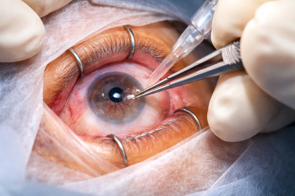
The Metal-in-Eye Removal Procedure

This is the part patients and families usually want to understand most. The truth is that there is no single technique that fits every case.
If the metal particle is only on the corneal surface, treatment may be much simpler and may sometimes be done under magnification at the slit lamp. But once the object has penetrated inside the eye, removal is usually performed in the operating theatre by an ophthalmic surgeon.
The procedure may involve:
- Closing any entry wound in the eye wall
- clearing blood or damaged vitreous
- precisely locating the foreign body
- removing it using microsurgical instruments
- repairing retinal tears or retinal detachment if present
- treating associated lens damage or traumatic cataract
- placing medication inside or around the eye when needed
So, the operation is often not only about removing the fragment. It is also about repairing injury, reducing infection risk, and preventing further complications.
What If the Retina Is Involved?
This makes the injury more serious.
If the metal fragment reaches the back of the eye, it can damage the retina or the vitreous. In such cases, vitrectomy is often needed so the surgeon can safely access the object and repair the surrounding damage. If there is a retinal tear or detachment, this usually needs to be treated at the same time. The final visual result then depends not only on removing the object, but also on how much damage occurred before treatment.
This is why early diagnosis and urgent treatment can make such a major difference.
Treatment Does Not End With Surgery
Patients sometimes think that once the metal is removed, the problem is over. In reality, intraocular foreign body treatment often continues well beyond the operation itself.
Ongoing care may include:
- antibiotic treatment
- anti-inflammatory medicines
- pressure monitoring
- retinal follow-up
- management of a traumatic cataract
- watching for scarring or infection
- additional surgery later if needed
In some patients, the first operation is only the beginning of recovery. That does not always mean the outlook is poor. It simply means that serious eye trauma can be complex and requires careful follow-up.
Recovery After Surgery
Recovery varies from one case to another. A small fragment treated quickly is very different from a deep penetrating injury with retinal damage.
After surgery, patients may experience:
- Redness and discomfort in the early phase
- blurred vision for some time
- The need for frequent eye drops
- several follow-up visits
- temporary activity restrictions
The final visual outcome depends on where the foreign body landed, whether infection developed, whether the retina or lens was injured, and how quickly treatment was started. Reviews of intraocular foreign body injuries consistently show that the severity of the original injury is one of the most important factors affecting visual recovery.
When Is This an Emergency?

A suspected intraocular foreign body should always be treated as urgent. This is not something to wait on overnight or manage with over-the-counter drops.
Seek immediate specialist eye care if:
- metal struck the eye at high speed
- Vision dropped suddenly after the injury
- The eye is painful and sensitive to light
- There is blood inside the eye
- Something appears to have penetrated the eyeball
- The pupil looks irregular
- You notice flashes, floaters, or a dark shadow in your vision
The safest rule is simple: if the history sounds serious, treat it seriously.
What Not to Do After a Penetrating Eye Injury
The negative first reaction can make the situation worse.
Do not:
- Rub the eye
- Press on the eyelid
- Try to remove the object yourself
- Use tweezers, cotton buds, or other tools
- Put random drops into the eye
- Delay treatment because the fragment seems small
Even a well-meaning attempt to help can push the object deeper or increase the risk of infection.
Can Vision Return to Normal?
Sometimes yes, but not always.
If the metal is removed quickly and the deeper structures of the eye are not badly damaged, recovery may be very good. If the retina, optic nerve, or central vision area has been injured, the final result may be more limited. The aim of treatment is always to save as much vision as possible, prevent infection, and keep the eye structurally stable.
For patients, that means one very important thing: getting the best possible chance of seeing clearly again.
FAQs
1. Is every metal fragment in the eye an intraocular foreign body?
No. Some fragments remain on the surface of the cornea or conjunctiva, while others penetrate deeper and enter the eye. The difference is extremely important and must be assessed by an eye specialist.
2. Will removal always happen on the same day?
Not every case follows the same timeline, but suspected penetrating eye injuries are treated urgently. The treating ophthalmologist decides the timing based on the condition of the eye and the extent of damage.
3. Is the surgery painful?
The procedure is usually performed under appropriate anaesthesia, so patients are kept comfortable. Some discomfort afterwards is normal and is managed with medication and follow-up care.
4. Can I drive home after treatment?
Usually, patients should not plan to drive themselves home. Vision may be reduced, and the eye may need protection or patching after treatment.
5. Can safety glasses really prevent this?
In many work-related and DIY cases, yes. Proper protective eyewear can make a major difference, especially during grinding, drilling, welding, or hammering metal.
Conclusion
A metal fragment inside the eye is never something to ignore. Intraocular foreign body removal is a highly specialised procedure that focuses not only on taking the fragment out, but also on repairing damage, preventing infection, and protecting vision. The earlier the injury is assessed, the better the chance of saving sight and avoiding long-term complications.
For urgent assessment and expert eye care in Dubai, patients who suspect a penetrating eye injury should seek immediate specialist attention without delay. To consult Dr Mandeep Lamba at Prime Hospital, Al Garhoud, Dubai, call +971 524 227 000 for urgent eye assessment.


