
Glaucoma can develop without pain, redness or an obvious change in eyesight. A person may continue working, driving and reading normally while the disease gradually damages the optic nerve. By the time a noticeable vision problem appears, some of the damage may already be permanent.
This is why glaucoma care is not only about treating symptoms. It is about identifying risk early, examining the optic nerve and monitoring changes before they begin to affect daily life. Although glaucoma cannot always be prevented, timely screening and appropriate glaucoma treatment in Dubai can often slow its progression and help protect the vision that remains.
For people with a family history of glaucoma, diabetes, high eye pressure or other risk factors, waiting for symptoms is not a safe approach. A comprehensive eye examination can identify changes that a routine vision test or glasses check may miss.
What Is Glaucoma?
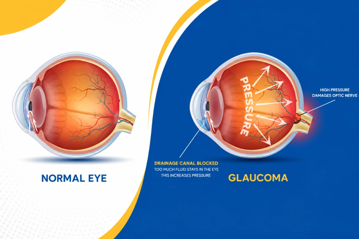
Glaucoma refers to a group of eye conditions that damage the optic nerve. The optic nerve carries visual information from the eye to the brain, making it essential for normal vision.
In many patients, glaucoma is associated with raised pressure inside the eye, known as intraocular pressure or IOP. The front of the eye continuously produces a clear fluid called aqueous humour. This fluid normally drains through natural drainage channels. When it does not drain efficiently, pressure may build up and place stress on the optic nerve.
However, eye pressure is only one part of the condition. Some people develop glaucoma even when their eye pressure is within the statistically normal range. Others may have high eye pressure for years without developing optic nerve damage. This is why an eye-pressure reading alone cannot confirm or rule out glaucoma.
The most common types include:
Primary open-angle glaucoma
This is the most common form. It usually develops slowly and may not produce noticeable symptoms during its early stages. Peripheral or side vision is commonly affected first.
Normal-tension glaucoma
In normal-tension glaucoma, optic nerve damage develops even though the measured eye pressure is not particularly high. The ophthalmologist may still recommend lowering the pressure because doing so can help slow further damage.
Angle-closure glaucoma
Angle-closure glaucoma occurs when the drainage angle between the iris and cornea becomes narrowed or blocked. It may develop gradually, but an acute attack can cause a sudden and dangerous rise in eye pressure.
Secondary glaucoma
Secondary glaucoma develops due to another eye or health condition. Possible causes include eye inflammation, previous eye trauma, advanced diabetic eye disease, certain eye procedures and prolonged use of corticosteroid medicines.
Congenital or childhood glaucoma
This is a rare form related to abnormal development of the eye’s drainage system. It may be present at birth or develop during childhood.
Can Glaucoma Be Prevented?
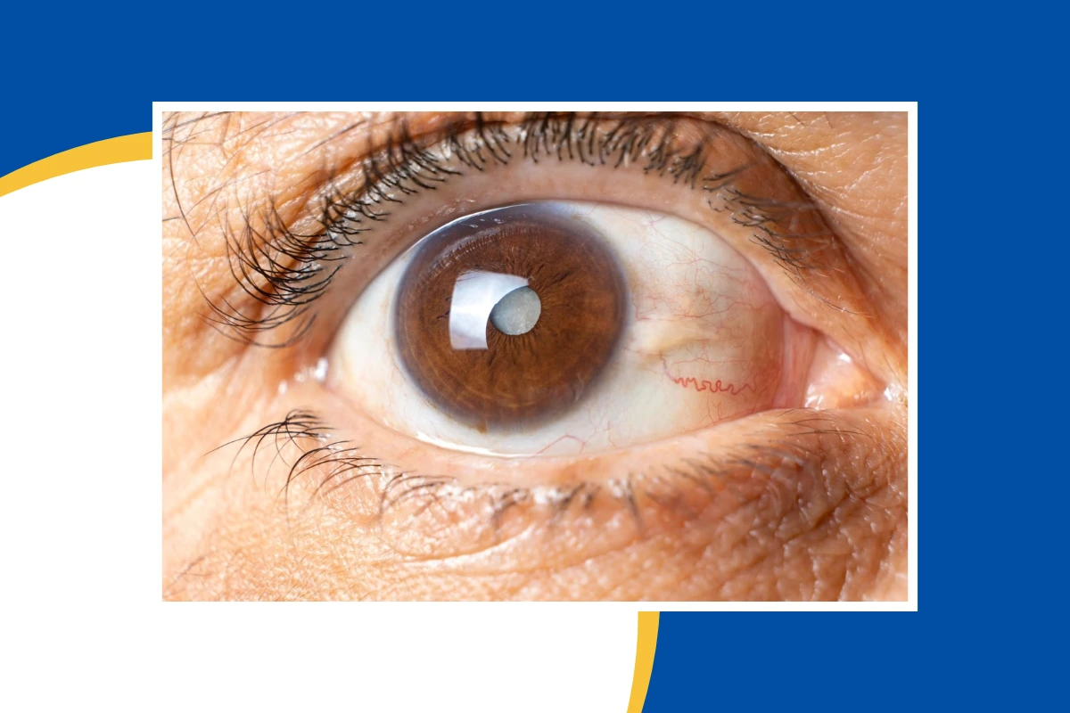
There is currently no guaranteed method of preventing every form of glaucoma. Ageing, inherited risk and the natural structure of the eye cannot be changed.
What can often be prevented or delayed is severe vision loss from undiagnosed or uncontrolled glaucoma.
Early detection allows an ophthalmologist to assess the optic nerve, establish a baseline and begin treatment when necessary. Lowering eye pressure is currently the main modifiable factor used to reduce the risk of progression. Treatment may involve monitoring, prescription eye drops, laser treatment or surgery, depending on the individual patient.
Therefore, prevention in glaucoma care mainly means:
- Detecting the condition before significant vision loss develops
- Identifying people who have ocular hypertension or suspicious optic nerves
- Monitoring high-risk patients regularly
- Controlling eye pressure when treatment is required
- Using prescribed medicines consistently
- Attending follow-up appointments even when the eyes feel normal
- Protecting the eyes from avoidable injuries
- Informing the ophthalmologist about steroid use and relevant medical conditions
The absence of symptoms does not mean the condition is stable. Glaucoma monitoring is based on clinical tests and comparison over time, not simply on how the patient feels.
Why Is Glaucoma Often Detected Late?
Most people expect an eye disease to cause blurred vision, discomfort or redness. Glaucoma often behaves differently.
During the early stages of open-angle glaucoma, central vision may remain clear. The brain can also compensate for small areas of missing peripheral vision, making early changes difficult to notice. According to the National Eye Institute and the CDC, around half of people with glaucoma may be unaware that they have it.
A patient may only begin noticing a problem after the visual field has become considerably restricted. Possible difficulties may include:
- Bumping into objects on one side
- Missing steps or kerbs
- Difficulty moving in dimly lit areas
- Trouble noticing vehicles or pedestrians approaching from the side
- Feeling less confident while driving
- Needing to turn the head more frequently to see surrounding objects
These changes can be gradual, which is why they are sometimes attributed to ageing, tiredness or a change in spectacle power.
What Are the Early Signs of Glaucoma?
For the most common form of glaucoma, there may be no clear early signs. This is the most important point for patients to understand.
A person can have normal reading vision and no eye pain while glaucoma is developing. Early glaucoma is usually detected during an eye examination rather than through symptoms.
As the disease progresses, a patient may experience:
- Gradual loss of side vision
- Patchy areas of missing vision
- Reduced ability to adjust in low light
- Difficulty navigating unfamiliar spaces
- Tunnel-like vision in advanced stages
- Loss of central vision in very advanced disease
These symptoms should not be used as a checklist for deciding whether to get tested. Waiting until they appear may mean waiting until some optic nerve damage has already occurred.
Emergency Symptoms That Should Not Be Ignored
Acute angle-closure glaucoma is different from the common slow-developing form. It can cause a sudden rise in eye pressure and requires immediate medical attention.
Seek urgent eye care or go to an emergency department if you develop:
- Sudden severe eye pain
- A red eye
- Sudden blurred vision
- Coloured rings or halos around lights
- A severe headache around the eye
- Nausea or vomiting with eye pain
- A rapid reduction in vision
Without urgent treatment, acute angle closure can cause serious and rapid damage to the optic nerve.
Who Is at Higher Risk of Developing Glaucoma?
Anyone can develop glaucoma, but certain factors increase the likelihood. Having one risk factor does not confirm that you have glaucoma. It means that regular and sometimes more detailed monitoring may be appropriate.
Increasing age
The risk of glaucoma generally increases with age. Adults over 60 are considered a higher-risk group, although glaucoma can occur earlier, particularly when other risk factors are present.
Family history
Glaucoma can run in families. Having a parent, sibling or child with glaucoma may increase your risk.
Patients should try to find out:
- Which relative has glaucoma
- At what age they were diagnosed
- Whether they required laser treatment or surgery
- Whether they experienced significant vision loss
Sharing this information with your ophthalmologist can help guide your screening plan.
High eye pressure
Raised intraocular pressure is one of the strongest recognised risk factors. However, one high reading does not automatically mean that a person has glaucoma.
Eye pressure can vary during the day and may be influenced by corneal thickness, medications, eye structure and other factors. The pressure result must be assessed alongside the optic nerve, visual field and imaging findings.
Diabetes
People with diabetes may have a higher risk of developing certain forms of glaucoma. Diabetes can also cause retinal disease, so regular dilated eye examinations are particularly important.
Steroid use
Steroid medicines can raise eye pressure in susceptible individuals. This can occur with steroid eye drops, tablets, injections, skin preparations or inhaled medicines.
Patients should not stop prescribed steroids without medical advice. Instead, tell your ophthalmologist which steroid you use, how frequently you use it and how long you have been taking it. Eye-pressure monitoring may be recommended.
Previous eye injury
An eye injury can damage the drainage system and lead to glaucoma months or even years later. A previous sports injury, workplace injury, road accident or blunt trauma should be mentioned during an eye consultation.
Previous eye inflammation or surgery
Conditions such as uveitis can affect eye pressure and drainage. Certain previous eye procedures may also change a patient’s glaucoma risk.
Eye structure and spectacle power
Some anatomical features may increase the risk of particular forms of glaucoma. For example, people with narrow drainage angles may be more susceptible to angle closure. Significant short-sightedness or long-sightedness may also be relevant, depending on the type of glaucoma being assessed.
Corneal thickness
The thickness of the cornea can affect the interpretation of eye-pressure measurements. It may also help the ophthalmologist assess a patient’s overall glaucoma risk.
Who Should Consider Glaucoma Screening in Dubai?
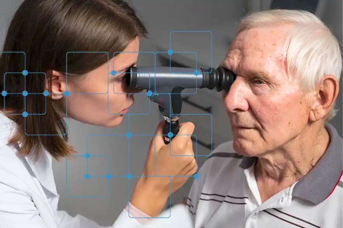
Glaucoma screening should be considered even when vision appears normal, especially when risk factors are present.
You may benefit from a comprehensive glaucoma assessment if you:
- Are over 40 and have never had a detailed eye examination
- Are over 60
- Have a parent, sibling or child with glaucoma
- Have previously been told that your eye pressure is high
- Have diabetes
- Use steroid medicines regularly or for prolonged periods
- Have experienced an eye injury
- Have had previous eye inflammation or surgery
- Have been told that your optic nerve looks suspicious
- Have significant short-sightedness or long-sightedness
- Notice changes in side vision
- Experience unexplained difficulty driving or seeing in low light
People at higher risk may need a comprehensive dilated eye examination every one to two years, although the correct interval should be decided by the treating ophthalmologist. Patients with diagnosed glaucoma or ocular hypertension may require more frequent monitoring.
What Happens During a Glaucoma Screening?
A proper glaucoma assessment involves more than a quick air-puff pressure test. No single test is sufficient for every patient.
The ophthalmologist may perform several of the following assessments.
Medical and family history
You may be asked about:
- A family history of glaucoma
- Diabetes and blood pressure
- Current medicines, including steroids
- Previous eye injuries
- Previous eye surgery
- Headaches, eye pain or halos
- Changes in driving or peripheral vision
Accurate information helps the ophthalmologist interpret the clinical findings.
Eye-pressure measurement
Tonometry measures the pressure inside the eye. It is usually quick and should not be painful.
A normal pressure reading does not completely exclude glaucoma, while a high reading does not automatically confirm it. The result must be interpreted in context.
Dilated optic nerve examination
Eye drops are used to widen the pupil so that the ophthalmologist can examine the retina and optic nerve more clearly.
The doctor looks for changes in the optic nerve’s shape, colour and nerve-fibre tissue. Dilated examination remains an important part of glaucoma assessment.
Optical coherence tomography
Optical coherence tomography, commonly called OCT, produces detailed scans of the optic nerve and retinal nerve-fibre layer.
The test can identify thinning that may be associated with glaucoma. It is also valuable for documenting a baseline and comparing future scans.
An OCT result should not be interpreted in isolation. The scan must be assessed alongside the clinical examination, visual field test, eye pressure and other findings.
Visual field test
A visual field test checks peripheral vision. During the test, the patient responds whenever a small light is seen in different parts of the visual field.
The test helps identify patterns of missing vision and monitor whether glaucoma is progressing. Since concentration and response timing can affect the result, the test may occasionally need to be repeated.
Gonioscopy
Gonioscopy allows the ophthalmologist to inspect the drainage angle of the eye. It helps determine whether the angle is open, narrow or closed and is important when assessing the risk of angle-closure glaucoma.
Corneal thickness measurement
Pachymetry measures the thickness of the cornea. This helps the ophthalmologist interpret the eye-pressure reading more accurately and assess overall risk.
What Happens If the Tests Suggest Glaucoma?
Not every suspicious result means that treatment must begin immediately.
Some patients are classified as glaucoma suspects. This may be because they have:
- High eye pressure without confirmed nerve damage
- An optic nerve that appears unusual
- A borderline visual field result
- A family history combined with other risk factors
- A narrow drainage angle
- Changes that require comparison over time
The ophthalmologist may recommend repeat pressure checks, OCT scans, visual field tests or photographs of the optic nerve before deciding whether treatment is necessary.
The aim is to avoid both extremes: ignoring genuine disease and treating a patient unnecessarily. A personalised monitoring plan is therefore an important part of glaucoma treatment in Dubai.
Glaucoma Treatment in Dubai: What Options Are Available?
Treatment is designed to lower eye pressure and reduce the risk of further optic nerve damage. It cannot restore nerve tissue or vision that has already been permanently lost.
The treatment plan depends on:
- The type of glaucoma
- The level of eye pressure
- The amount of existing nerve damage
- The speed of progression
- The patient’s age and general health
- Corneal thickness
- Previous eye procedures
- The ability to use eye drops correctly
- The target pressure required to protect the optic nerve
Prescription eye drops
Eye drops are commonly used to lower eye pressure. Some improve fluid drainage, while others reduce the amount of fluid produced inside the eye.
These medicines usually need to be used consistently every day. They may not make the vision feel clearer because their purpose is to protect against future loss rather than create an immediate improvement.
Patients should tell their ophthalmologist if they experience:
- Burning or persistent redness
- Eyelid irritation
- Changes around the eye
- Breathing difficulty
- Changes in heartbeat
- Tiredness or dizziness
- Difficulty following the prescribed schedule
Do not stop glaucoma drops without discussing the problem with your doctor. A different medicine, dosage or treatment method may be available.
Laser treatment
Laser trabeculoplasty may be used for certain forms of open-angle glaucoma. The laser is applied to the eye’s drainage area to help fluid drain more effectively and reduce pressure.
The procedure is usually performed in the clinic with numbing drops. Some patients may still need eye drops afterwards, and the effect of laser treatment can reduce over time.
A different laser procedure, such as laser peripheral iridotomy, may be recommended for patients with narrow angles or angle-closure risk. It creates a small opening in the iris to improve fluid movement and reduce the risk of pressure blockage.
When Is Glaucoma Surgery in Dubai Considered?
Glaucoma surgery in Dubai may be considered when medicines and laser treatment do not reduce eye pressure sufficiently, when glaucoma continues to progress or when the pressure needs to be lowered more significantly.
Surgery may also be discussed when:
- Glaucoma is already advanced
- The patient cannot tolerate prescribed eye drops
- Using multiple drops has become difficult
- Eye pressure remains above the target level
- The optic nerve or visual field continues to worsen
- The type of glaucoma is unlikely to be controlled with drops alone
The recommendation is based on the risk of further vision loss, not merely on one pressure reading.
Trabeculectomy
Trabeculectomy creates a new pathway through which fluid can drain from the eye. It can achieve a significant reduction in pressure and may be considered for patients with more advanced or difficult-to-control glaucoma.
Glaucoma drainage implant
A small tube or drainage device is placed in the eye to help fluid drain and lower pressure. This may be used for several forms of complex or secondary glaucoma.
Minimally invasive glaucoma surgery
Minimally invasive glaucoma surgery, commonly known as MIGS, includes a range of procedures designed to lower pressure with smaller incisions and, in selected patients, a faster recovery.
MIGS may be suitable for some patients with mild or moderate glaucoma and is sometimes performed alongside cataract surgery. It is not automatically the best option for every patient, particularly when a very low target eye pressure is required.
Glaucoma surgery cannot reverse existing vision loss or permanently cure the disease. Its purpose is to lower pressure and protect the remaining vision. Continued follow-up is necessary even after a successful procedure.
Can Lifestyle Changes Prevent Glaucoma?
No food, vitamin, exercise or home remedy has been proven to replace glaucoma screening or prescribed treatment.
General healthy habits remain valuable for overall eye and cardiovascular health. These may include:
- Keeping diabetes and blood pressure well controlled
- Exercising regularly after discussing any restrictions with your doctor
- Avoiding smoking
- Eating a balanced diet
- Wearing protective eyewear during sports or hazardous work
- Attending routine medical and eye appointments
- Informing doctors about all medicines and supplements being used
Patients should be cautious about supplements advertised as glaucoma cures. Glaucoma requires clinical monitoring, and delaying effective care in favour of unproven remedies can allow irreversible damage to continue.
How Can Patients Protect Their Vision After a Glaucoma Diagnosis?
A glaucoma diagnosis can feel worrying, but many patients maintain useful vision for years with appropriate treatment and regular monitoring.
The following steps can make a meaningful difference:
Use your drops at the correct time
Connect your eye drops with a regular activity, such as brushing your teeth or having a meal. Phone reminders can also help.
Learn the correct eye-drop technique
Place one drop into the eye without touching the bottle tip to the eyelid or lashes. Gently closing the eye and pressing the inner corner for a short time may reduce the amount of medicine entering the nose and bloodstream.
Attend follow-up appointments
Glaucoma treatment cannot be judged by symptoms alone. Your doctor needs to repeat pressure measurements, optic nerve imaging and visual field tests to determine whether the disease is stable.
Report side effects
Do not silently tolerate a medicine that causes significant discomfort or systemic symptoms. Discuss it with your ophthalmologist rather than stopping treatment independently.
Keep a record of your medicines
Bring an updated list of eye drops, tablets and inhalers to appointments. This is especially helpful when consulting more than one doctor.
Encourage relatives to get tested
Since glaucoma can run in families, close relatives should be informed and encouraged to have comprehensive eye examinations.
The Most Effective Prevention Is Early Detection
Glaucoma cannot always be prevented from developing, but severe vision loss should not be accepted as unavoidable. The most useful protection is to understand your risk, have a comprehensive eye examination and follow an appropriate monitoring or treatment plan.
Do not wait for pain or blurred vision before arranging an assessment. Most early glaucoma does not announce itself through symptoms.
For patients looking for personalised glaucoma treatment in Dubai, Dr Mandeep Lamba can assess eye pressure, optic nerve health, visual fields and individual risk factors before recommending monitoring, medication, laser treatment or glaucoma surgery in Dubai where clinically appropriate. Early evaluation offers the best opportunity to protect your remaining vision and maintain confidence in everyday activities.
Frequently Asked Questions About Glaucoma
1. Can glaucoma be completely cured?
Glaucoma is generally managed as a long-term condition rather than permanently cured. Treatment can lower eye pressure and slow or stop further damage, but it cannot restore optic nerve tissue that has already been lost.
2. Can I have glaucoma with normal vision?
Yes. Central vision can remain clear during the early stages, while subtle damage develops in the optic nerve or peripheral visual field.
3. Does normal eye pressure mean I do not have glaucoma?
No. Normal-tension glaucoma can develop without a pressure reading above the usual statistical range. Diagnosis requires assessment of the optic nerve, visual field and other clinical findings.
4. Does high eye pressure always mean glaucoma?
No. Some people have ocular hypertension without detectable optic nerve damage. However, they may require monitoring or treatment depending on their overall risk.
5. Can glasses correct vision lost from glaucoma?
Glasses can correct refractive errors such as short-sightedness, long-sightedness and astigmatism. They cannot restore vision that has been permanently lost because of optic nerve damage.
6. Will every glaucoma patient need surgery?
No. Many patients are managed with monitoring, eye drops, laser treatment or a combination of these. Glaucoma surgery in Dubai is recommended when the expected benefit outweighs the risks and when other methods are not providing sufficient control.
7. Can glaucoma return after surgery?
Surgery lowers eye pressure but does not remove the underlying tendency to develop glaucoma. The effect may change over time, and some patients may need medicines, laser treatment or another procedure later.
8. How often should I be screened?
The interval depends on your age, family history, eye pressure, optic nerve appearance and other risk factors. Higher-risk patients may be advised to have a comprehensive dilated examination every one to two years, while diagnosed patients usually require more frequent monitoring.


